
Ureteroscopy (rigid and flexible)
Accessing calculation through natural means
Discover flexible or rigid ureteroscopy for treating urinary stones naturally. Dr. Sébastien Dominique, urologist, offers a precise and minimally invasive treatment.
Exploration and extraction of urinary stones via the natural route
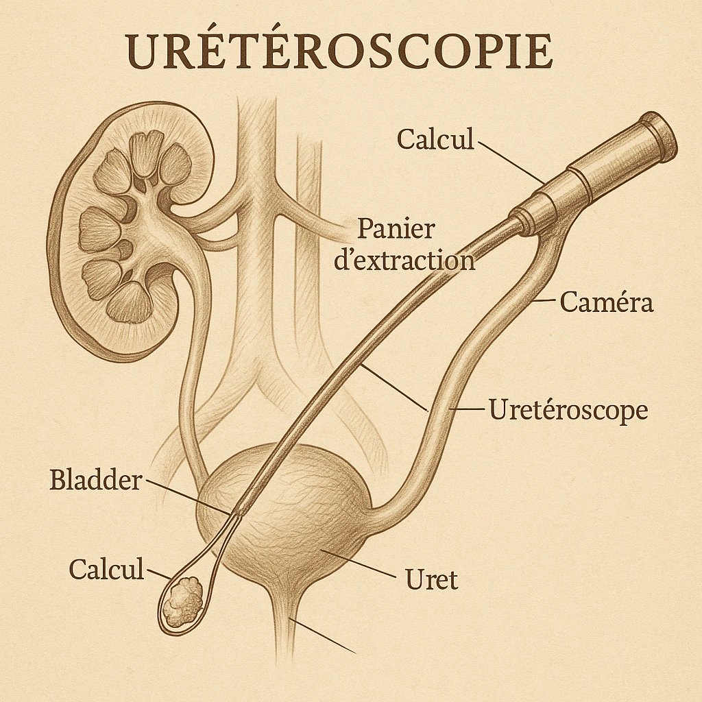
What is ureteroscopy (rigid or flexible)?
Ureteroscopy is an endoscopic urological procedure that allows access to the upper urinary tract (ureter, renal pelvis, calyces of the kidney) through the natural passages —without surgical incision. There are two types:
- Rigid ureteroscopy : used mainly for stones located in the ureter (pelvic segment, iliac…), or to extract fragments, place probes, etc.
- Flexible ureteroscopy : allows navigation into the cavities of the kidney, including the calyces, thanks to a flexible endoscope, thinner and more malleable, adapted to anatomical curves.
The main objective is the direct exploration of the excretory system, the visualization of the stone and then its fragmentation (most often with a laser) or its extraction (forceps, basket) via these natural pathways, without opening the skin.
Ureteroscopy Expertise

In what cases is this intervention offered?
Main indications
Ureteroscopy, rigid or flexible, is indicated particularly in the following situations:
- Urinary stones that are resistant or difficult to access with extracorporeal shock wave lithotripsy (ESWL), especially dense stones (> ~ 1000 Hounsfield units) or hard stones (e.g. cystine stones).
- Moderately sized stones (< ~ 20 mm) in the ureter or kidney, when ESWL is not effective or possible.
- Renal localization, particularly in the lower calyces or in malformed kidneys, or when the kidney is difficult to access for lithotripsy.
- An alternative to ESWL if it is contraindicated (e.g., patient on anticoagulants, unfavorable anatomy, high stone density).
Limitations and contraindications
- Untreated urinary tract infection.
- Difficult ureteral anatomy (stenosis, history of ureteral surgery, abnormalities of the path, very large prostate) which prevents access or passage of the device.
- Very large stones (> ~ 20-25 mm), or staghorn or very complex stones, for which other techniques (percutaneous nephrolithotomy) will be preferable.
How does the procedure unfold?
Here are the typical steps of flexible or rigid ureteroscopy for the exploration and extraction of urinary stones:
Preparation
- Imaging assessment (CT scan without injection, ultrasound, possibly X-ray) to precisely locate the stone, measure its size, and evaluate its density.
- Urine culture (ECBU – cytobacteriological examination of urine) to check sterility or treat an infection before the procedure
- Anesthesia: often general for flexible, sometimes local or spinal anesthesia depending on the patient’s condition and the location of the stone.
- Sometimes a double-J stent is inserted beforehand to dilate the ureter (“prepare” the passage of the ureteroscope), if necessary.
Intervention
- The urethroscope is introduced through the urethra, then the bladder, and then into the ureter; for the flexible version, progression is made to the renal cavities if necessary.
- Visualization of the calculation, fragmentation (most often Holmium YAG laser) if too large to be extracted directly.
- Extraction of fragments or the entire stone using forceps or an endoscopic basket.
- At the end of the procedure, a double-J stent (ureteral stent) is most often inserted to drain the stent, prevent edema, facilitate the removal of fragments, or relieve pain.
Post-operative care
- Short hospitalization, often on an outpatient basis or 24 hours depending on the case.
- Moderate pain, possible transient urinary bleeding, sometimes discomfort related to the double-J stent (pulling, frequent urges).
- Pain relievers, possibly anti-inflammatories prescribed.
- Monitoring of kidney function, urine monitoring.
- Removal of the double-J stent according to protocol (a few days to a week depending on tolerance and the site of the stone).
- Radiological or imaging control a few weeks later, to ensure the absence of residual stone fragments.
Expected results
- A high success rate for pelvic ureter stones, often > 90%.
- For moderately sized kidney stones (< ~1 cm), good results; for sizes between 1-2 cm, less success, and sometimes the need for several interventions or additional treatment.
Possible complications
- Transient hematuria (bleeding in the urine) after the act.
- Pain or discomfort (colic, clot, ureteral spasm) in the days that follow.
- Urinary tract infection, or even more severe if sampling or fragmentation occurs with a pre-existing, untreated infection.
- Risk of ureteral injury (perforation, laser burn, avulsion, or trauma) to be handled with care.
- Distant ureteral stenosis in some cases.
Expertise
Kidney stone: scarless treatment by flexible ureteroscopy (uncensored) – Dr. Dominique

Why choose this technique rather than an incision or other method?
Pre- and post-operative
Pre-operative: what you need to know
- The patient will need to undergo tests before the procedure: imaging (non-contrast CT scan, ultrasound) to locate the stone, measure its density and size.
- Checking for the absence of a urinary tract infection (urine culture), and treatment if necessary.
- Information on anesthesia, hospitalization procedures (outpatient or 1 day depending on the facility).
- Review of current treatments (anticoagulants, antiplatelet agents), possible adjustment.
- Discussion on the possible placement of a preliminary probe (double-J) if the ureter is not sufficiently dilated for the passage of the endoscope.
Post-procedure follow-up
- Moderate pain is expected, slight bleeding is possible, often lasting a few days.
- Take painkillers/anti-inflammatories as needed.
- Drink plenty of fluids to help eliminate stone fragments.
- In the case of a double-J stent, it will be removed according to protocol (a few days to a week or more depending on the situation).
- A radiological/imaging check a few weeks later to ensure that no fragments remain.
- Urological monitoring to monitor for possible complications, including stenosis, infection, and discomfort related to the catheter.
Experience
Is it possible to play sports with a double J stent?

Specialist in ureteroscopy
My review in video
I made this video to explain flexible or rigid ureteroscopy in detail.
Make an appointment
Dr. Sébastien Dominique offers you a personalized assessment, an appropriate procedure, and careful follow-up to guarantee the best possible result.
FAQ – Frequently Asked Questions
What is a ureteroscopy?
This involves examining the ureter and kidney using a thin endoscope inserted through the natural openings, without incision. The rigid ureteroscope treats the pelvic ureter; the flexible ureteroscope allows access to the kidney and all its cavities.
In what cases is it indicated?
Primarily for fragmenting and extracting urinary stones with lasers, but also for investigating hematuria, biopsying lesions of the excretory tract, or treating certain ureteral strictures.
Is the procedure painful?
It is performed under general anesthesia and is not painful. Afterwards, there is usually only temporary burning during urination and the possible discomfort of a temporary JJ stent.
Why is a JJ stent sometimes inserted?
The JJ stent, placed between the kidney and the bladder, protects the ureter and facilitates urine flow after the procedure. It is removed during a follow-up appointment after a few days to a few weeks, depending on the case.
Dr Sébastien Dominique's expertise
Chirurgien Urologue à Paris

Ureteroscopy (rigid and flexible): there are solutions.