
Urinary stones (urolithiasis)
Understand the origin of your calculations and take action
Prevent the recurrence of urinary stones with advice from Dr. Dominique: hydration, diet, metabolic assessment and personalized follow-up.
Introduction
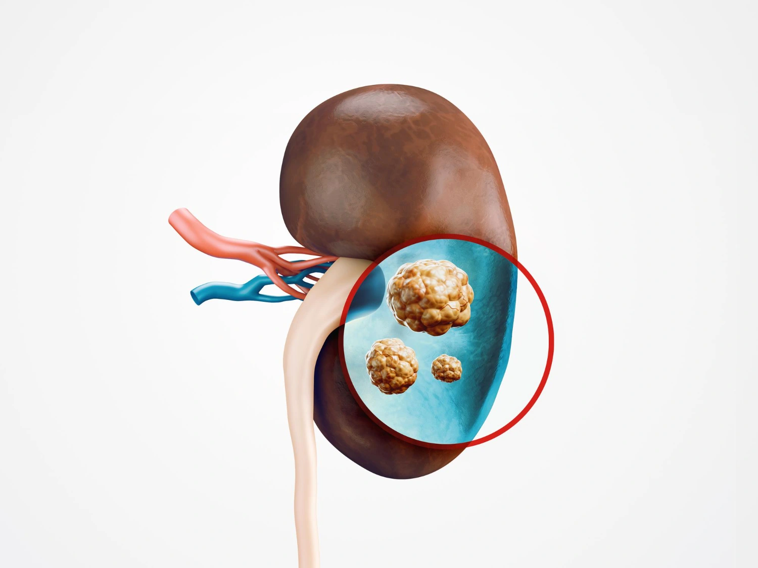
Urinary lithiasis, or “kidney stones,” refers to the formation of small crystals or stones in the urinary tract (kidney, ureter, bladder). These stones can be asymptomatic or become very symptomatic when they move or obstruct the flow of urine.
Dr. Dominique specializes in the diagnosis, minimally invasive treatment, and prevention of recurrence in urology.
If a renal colic attack occurs or blood appears in the urine, prompt consultation is recommended to avoid kidney or infectious complications.
Symptoms and warning signs
Diagnosis
Care and treatment
Experience
Expertise in recurrent kidney stones
Prevention of relapse
An essential aspect of treatment is prevention in order to limit recurrences:
- Lithogenic metabolic assessment : analysis of urine and blood to identify risk factors (hypercalciuria, hyperoxaluria, hyperuricosuria, inhibitor deficiency, etc.).
- Dietary adjustment :
- Increased water consumption (≥ 1.5 to 2 L/day depending on tolerance)
- Moderation of salt intake
- Control of animal protein and dietary oxalate intake according to context
- Adjusting calcium intake (avoiding excesses, but also deficiencies)
- Prophylactic drug treatment according to the type of stone (thiazides, citrates, allopurinol, etc.).
- Regular monitoring : control imaging, urological and metabolic follow-up.
Why consult a specialist urologist?
How to prevent the recurrence of urinary stones?
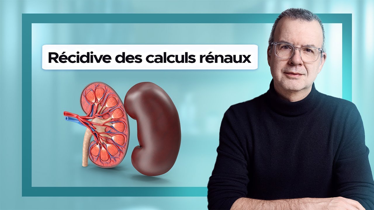
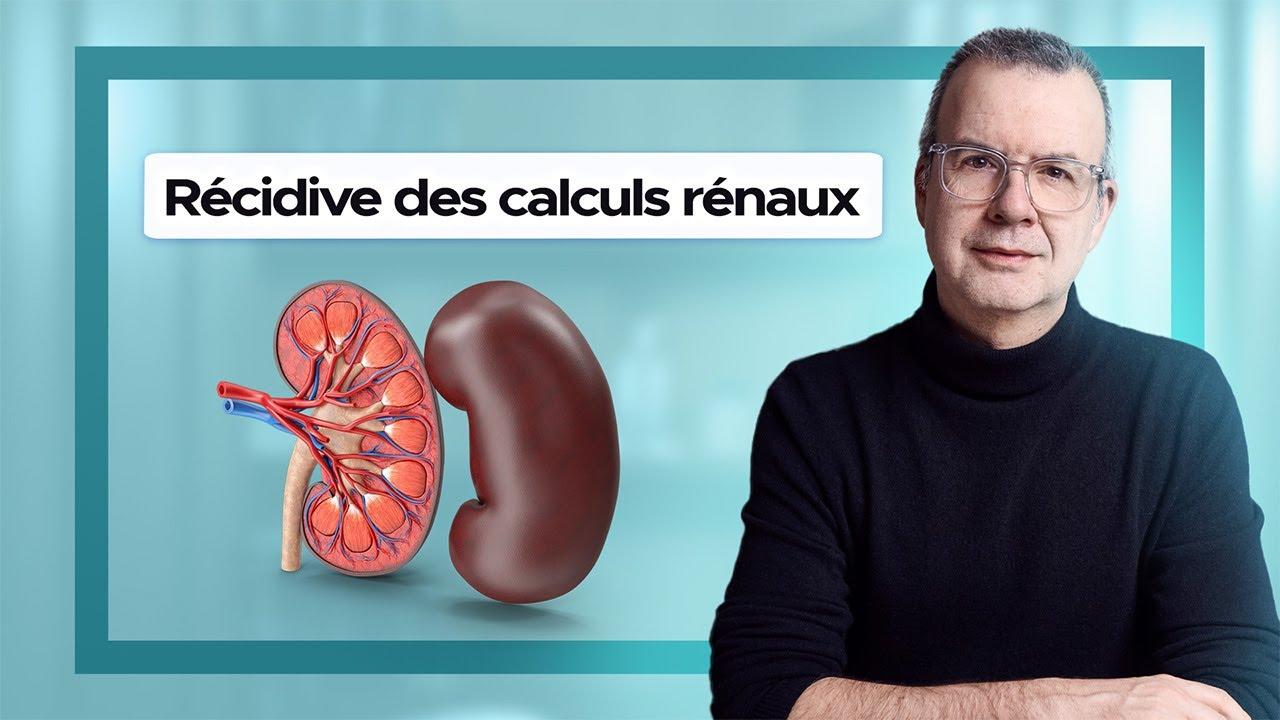
In this video, I explain how to effectively reduce the risk of recurring kidney stones. I share essential daily habits to adopt, mistakes to avoid, and the importance of a metabolic assessment to understand the origin of your stones. My goal: to help you prevent another attack and protect your kidney health in the long term.
Do you suffer from urinary stones?
Dr. Sébastien Dominique provides emergency care for kidney stone attacks , as well as follow-up care for patients prone to kidney stones . Urology Practice Paris Opéra – Paris 17th. Consultations available in person or via teleconsultation. Book your appointment today.
FAQ – Frequently Asked Questions
How do urinary stones form?
Kidney stones form when urine is too concentrated in certain mineral salts (most often calcium oxalate, uric acid, phosphates). Insufficient hydration, an unbalanced diet, and metabolic or familial predispositions are the main contributing factors.
What are the signs of a urinary stone?
The most typical sign is renal colic: intense lower back pain radiating to the groin. A kidney stone can also manifest as blood in the urine, recurrent infections, or remain asymptomatic for a long time and be discovered through imaging.
What tests allow for diagnosis?
A non-contrast abdominal and pelvic CT scan is the gold standard: it locates the stone and measures its size and density. Ultrasound is useful as a first-line investigation and in pregnant women. Blood and urine tests complete the diagnosis.
Can a kidney stone be eliminated without intervention?
Yes: most kidney stones smaller than 6 mm pass spontaneously, aided by adequate hydration and treatment to facilitate expulsion. For stones larger than 6 mm, or in cases of uncontrollable pain, infection, or kidney blockage, urological treatment is necessary.
Dr Sébastien Dominique's expertise
Chirurgien Urologue à Paris

Urinary stones (urolithiasis): there are solutions.